Inside the ear
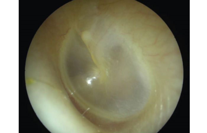
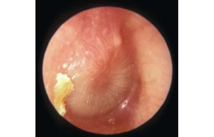
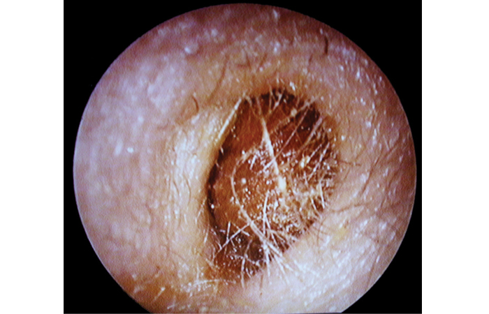
When using an otoscope, a normal eardrum (Figure 1) looks translucent and greyish in colour. Acute otitis media causes inflammation of the eardrum (Figure 2). The main impediments to seeing the eardrum clearly are debris or inflammation of the ear canal (usually due to otitis externa), or wax. Wax appears as a yellowy, orange or brown deposit (Figure 3).
Excluding the presence of acute infection is important and is mostly achieved through careful history taking. Otoscopy provides additional information and in acute otitis media (AOM) a distinctly red, yellow, or cloudy eardrum (tympanic membrane) is usually seen (Figure 2). There may also be moderate to severe bulging of the eardrum, with loss of normal landmarks and an air-fluid level behind the drum which indicates a middle ear effusion. In severe cases there may be perforation of the eardrum and/or discharge in the ear canal.
OE appears as a red, sometimes swollen ear canal with potential debris (desquamated skin) or discharge, indicating inflammation and irritation of the ear canal, often accompanied by narrowing due to the swelling. The eardrum may be partially obscured by the swelling but should generally appear normal if the infection is confined to the external ear.
Identification aid
A left normal ear drum.
Appearance of eardrum in acute otitis media – inflammation and loss of landmarks. A small amount of yellow earwax is at bottom left.
Otoscopy of excess earwax.
Reflective exercise
Which preparations for earwax have you found o be most frequently requested by patients? How do these compare with your usual, personal recommendations?