Our ears help with both hearing and balance, but they are often taken for granted €“ until something goes wrong. In such situations, customers may turn to the pharmacy for help.
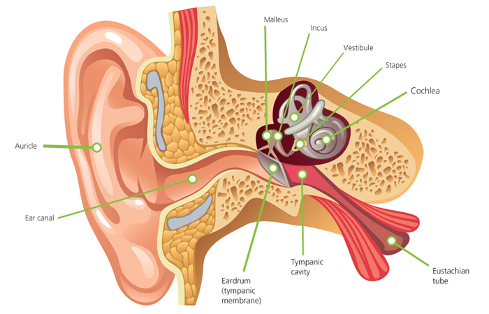
To understand the different conditions that can affect the ears, it is useful to know how the ear is structured. Broadly speaking, the ear is comprised of three distinct sections €“ the outer, the middle and the inner ear.
The outer ear consists of the auricle (pinna), which is made up of flexible cartilage that extends into the ear canal. At the far end of the ear canal is the eardrum (tympanic membrane), which sits between the outer and the middle ear.
The middle ear is an air-filled space. It contains the ossicles €“ three small bones that connect the eardrum to the inner ear and transmit vibration. These bones are called the malleus, the incus and the stapes, but are more commonly known as the hammer, anvil, and stirrup. The eardrum is a small circle of skin, comprised of three layers. The inner layer of the eardrum is made up of cells that produce mucus when irritated (e.g. by smoke or infection). The eustachian tube connects the middle ear to the back of the throat and helps to drain fluid and keep pressure within the ear at the right level. Changes in pressure (e.g. when flying) can cause pain. This tube is shorter in children, so they are more at risk of ear infections.
The inner ear consists of the cochlea, a fluid-filled, snail-shaped organ that makes hearing possible by converting sound into electrical pulses; the vestibular system (situated in the vestibule), which plays a key role in balance; and the vestibular and auditory nerves, which connect the inner ear to the brain.